
![iconzebrafish[1]](https://biolink-research.com/wp-content/uploads/2025/03/iconzebrafish1.png)

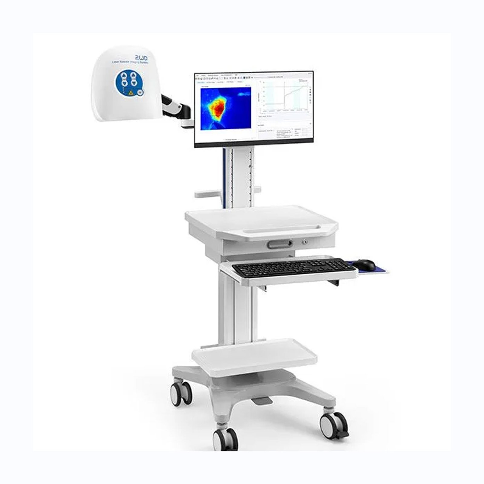
RFLSI-ZW – Laser Speckle Contrast Imaging System
RFLSI-ZW laser speckle imaging system is an even better tool for microcirculation research based on laser speckle contrast imaging technology (LSCI).
Laser Speckle Contrast Imaging System
RFLSI-ZW laser speckle imaging system is an even better tool for microcirculation research based on laser speckle contrast imaging technology (LSCI).
With the advanced optical design and improved image processing algorithm, RFLSI-ZW Laser Speckle Contrast Imaging System shows greater performance in imaging field size, image quality, full-field frame rate and optical resolution, and provides a powerful and efficient means for human and animal tissue blood flow measurement.
Overview
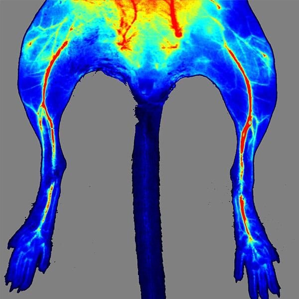
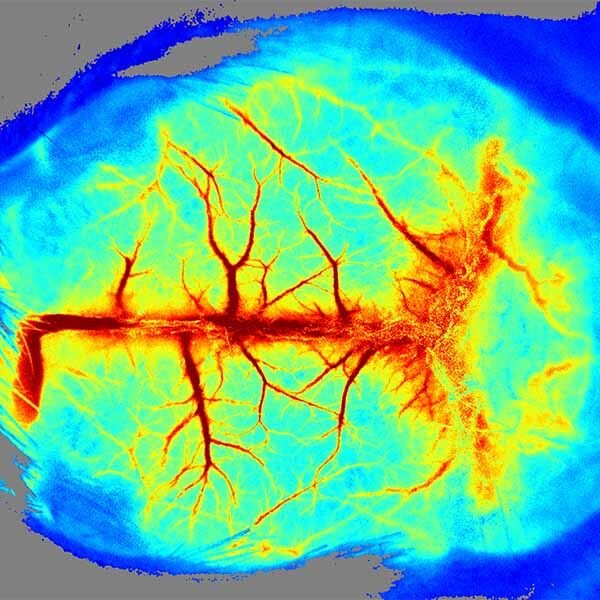
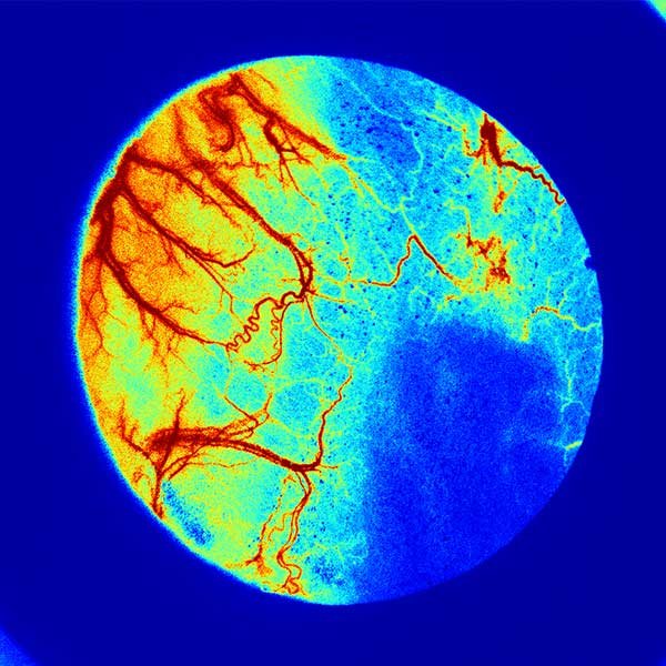
The LSCI technology advantages are its non-contact, no contrast agent required, high frame-rate, high spatial resolution. They can be used to observe and record blood perfusion of any exposed tissues or organs for microcirculation study or pre-clinical researches like ischemic stroke, lower limbs, mesentery, etc. Multi-Output includes blood perfusion images and videos (500+ million pixels), quantified data for perfusion unit and vessel diameter.
Since 2019, our imaging system has been adopted by more than 200 colleges, universities, and research institutes worldwide such as Stanford University School of Medicine, Yale University, University of Manchester, Duke university, University College London, University of Tasmania, Universitaet Gesamthochschule Essen, Korea University. What’s more, it has contributed to publishing more than 200 reputed research papers in magazines like Nature Neuroscience、Gut、Brain、Blood、Circulation Research、Nano Today、Nature Communications、 Advanced Functional Materials and Diabetes.
Application
- Image any exposed tissue (skin or surgically exposed tissues) and species.
- Non-contact, non-contrast agent depending measurement.
- The built-in CMOS global shutter camera can achieve faster data acquisition and processing speed.
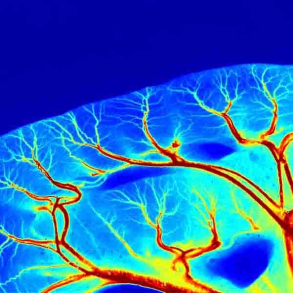
- Best optical resolution of 3.9 μm/pixel, providing more detailed tissue structures.
- Max frame rate (full field) up to 100 fps, acquiring real-time changes in larger areas.
- Motorised 10x optical zoom and auto focus. Image size ranges from 0.57×0.75 to 22.5×30 cm2 in all-in-one imager, covering multiple research applications.
- Fast auto and fine manual focus, improving focus efficiency and accuracy on various tissues.
- Optimal lens assembly, filtering the ambient and reflecting light.
- Class 1 of measurement and indicating lasers, safe to use without eye protection System.
- Laser stability hardware for the ultimate in reliable and consistent measurement over minutes, hours and days.
- Calibration with calibration box. Self-calibration is possible at any time to keep the equipment in optimal working condition.
- Trigger In/Out BNC connections for communication with external devices.
- Unlimited installation of analysis software in PC.
Highlights Of RFLSI-ZW Laser Speckle Contrast Imaging System
- Faça a imagem de qualquer tecido exposto (pele ou tecidos expostos cirurgicamente) e espécies.
- Medição sem contato e sem contraste dependente do agente.
- A câmera com obturador global CMOS embutida pode obter maior velocidade de aquisição e processamento de dados.
- Melhor resolução óptica de 3,9 μm/pixel, fornecendo estruturas de tecido mais detalhadas.
- Taxa máxima de quadros (campo total) de até 100 fps, adquirindo alterações em tempo real em áreas maiores.
- Zoom óptico motorizado de 10x e foco automático. O tamanho da imagem varia de 0,57×0,75 a 22,5×30 cm2 em um gerador de imagens multifuncional, abrangendo várias aplicações de pesquisa.
- Foco manual rápido e automático, melhorando a eficiência e a precisão do foco em vários tecidos.
- Montagem ideal da lente, filtrando a luz ambiente e refletindo a luz.
- Classe 1 de lasers de medição e indicação, seguros para uso sem sistema de proteção ocular.
- Hardware de estabilidade do laser para obter o máximo em medições confiáveis e consistentes em minutos, horas e dias.
- Calibração com caixa de calibração. A autocalibração é possível a qualquer momento para manter o equipamento em condições ideais de funcionamento.
- Conexões BNC de entrada e saída de disparo para comunicação com dispositivos externos.
- Instalação ilimitada do software de análise no PC.
Publications
RWD Laser Speckle Contrast Imaging System has contributed to publishing more than 100 reputed research papers in journals like Nature Communications, Blood, Circulation Research, Brain, Diabetes, and Theranostic. The paper list we collect contains about 18 research topics such as Cerebral Ischemia & Ischemic Stroke, Traumatic Brain Injury (TBI), Angiogenesis, Diabetes, Alzheimer’s Disease, Wound Healing, Limb Ischemia, etc.
Advantages
HD image & Video
2064*1544 high-resolution images; Optimize imaging details through algorithmic image stacking.
Accurate Data
Precise temperature control; Self-calibration program; Multiple optical lens coatings.
Large imaging area
Image size ranges from 5.7mm x 7.5mm to 225mm x 300mm; Motorised 10x optical zoom and auto focus.
Fast imaging
A high-speed camera (up to 100 FPS); Fast data transfer with USB3.0.
Easy to use
No contrast agent required; Flexible stand and cart options; BNC communication interface.
Safe Laser
Class 1 per IEC 60825-1:2014 – Safe to use without eye protection.
Successful Cases
Journal: Nature Neuroscience
Use the Laser Speckle Imaging System (LSCI) to detect the CBF in mouse barrel cortex during whisker stimulation.
Journal: Circulation Research
Use the Laser Speckle Imaging System to measure blood flow in cerebral arteries via a cranial window in anesthetized mice, to corroborate an in vivo physiological role for pS1928 upon HG.
Journal: Brain
Use laser speckle imaging to corroborate haemodynamic changes within the targeted region on the ipsilateral side of the brain before and after stroke in the photothrombotic stroke model.
Clients Say
Clinical & Experimental Epilepsy
“I couldn’t suggest further hardware features as I feel everything the user would need is there. The quality of the images is wonderful, The pixel resolution of the images is very impressive also. The user friendly software allows the user to control all aspects of both the operation of the machine but also the analysis of the results. The features available for data analysis, such as the ROI function and blood perfusion graph, allow for exciting results ...

Dr Samuel M Flaherty
Stroke & Vascular Function
“We can use the Laser Speckle Imaging System to observe the 5th branch of Mesentery.”

Yi-Je (Jay) Chen, Ph. D.
Brain Injury
"The instrument is incredibly easy to use allowing high-resolution images that can be used to assessed cerebral blood perfusion and vessels diameter at a highly reproducible level. I found impressive the possibility to monitor in real-time the results of several ROIs using the perfusion graph function provided in the software."

Dr. Federico Moro
Our Customer
If you are a researcher in neuroscience, dermatology or other fields, RFLSI-ZW Laser Speckle Contrast Imaging System can help you tell the results you need.